Figure 5.
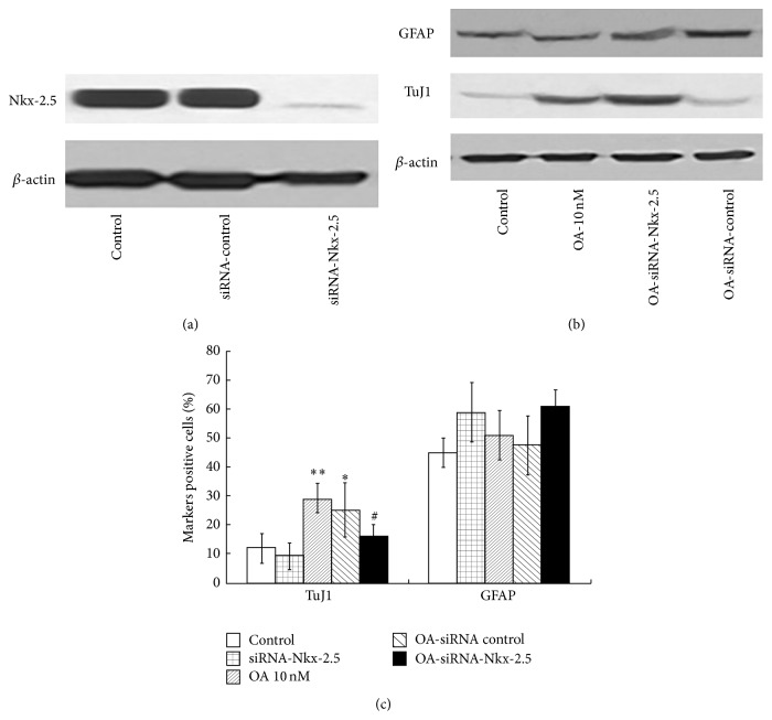
The siRNA of Nkx-2.5 was added to NSCs; 24 h later, cells were subjected to Western blotting analysis, indicating a marked knock-down of Nkx-2.5 expression (a). OA was added to the cells with silence of Nkx-2.5. The TuJ1 and GFAP protein expression was detected with Western blotting (b). Meanwhile, the immunofluorescence analysis of the percentage of TuJ1 and GFAP-positive cells was performed (n = 4). Results showed OA treatment resulted in significantly increase of the percentage of TuJ1-positive cells. However most effects of OA were abolished in OA-siRNA-Nkx-2.5 (n = 3; P < 0.05 versus OA 10 nM group), while the percentage did not significantly change in OA-siRNA-control (b and c). Scale bars: 100 μm. Results were expressed as mean ± SD from 4 independent experiments. * P < 0.05 ** P < 0.01 versus control; # P < 0.05 versus OA 10 nM.

