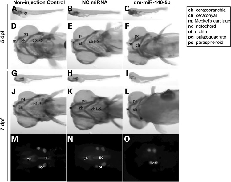
Figure 9.
Craniofacial and overall morphological phenotypes of zebrafish larvae in microRNA microinjection experiments. (A-O) Left, middle and right panels depict images corresponding to non-injection control, NC miRNA-injected and dre-miR-140-5p-injected groups at 5 dpf and 7 dpf larval stages, respectively. (A-E) 5 dpf stage of zebrafish larvae after microinjection with respective miRNAs were stained with Alcian blue 8 GX to observe overall morphological phenotypes (A-C) or craniofacial cartilage structures (D-E). (G-O) 7 dpf stage of zebrafish larvae after microinjection with respective miRNAs were stained with both Alcian blue 8 GX (G-L) and Alizarin red S (M-O) to observe overall morphological phenotypes (G-I) or craniofacial cartilage structures (J-L) or mineralized bone (MO). The RNA sequence of dre-miR-140-5p is exactly the same as that of hsa-miR-140-5p, indicating cross-species conservation (Supplementary Material, Fig. S1). Three independent experiments comparing non-injection control, NC miRNA-injected and dre-miR-140-5p-injected groups were performed, and consistent morphological phenotypes were obtained for each of these three groups across these experiments. Images presented are from one representative experiment. NC, negative control; dpf, days post-fertilization.

