Figure 2.
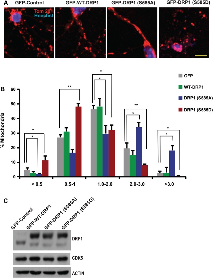
DRP1 phosphorylation at Ser585 regulates mitochondrial morphology in cerebellar granule neurons. (A) Cerebellar granule neurons (CGNs) were infected with adeno-associated viruses for GFP-expressing wild type DRP1 (GFP-WT-DRP1), GFP-expressing DRP1 mutants [GFP-DRP1 (S585A) and GFP-DRP1 (S585D)] or a GFP control for 6 days. GFP was fused to DRP1 constructs at the N terminal on the same promoter. Neurons were fixed and stained with TOM-20 antibody to assess mitochondrial morphology. Nuclei were stained with Hoechst. Representative images of mitochondrial morphology under different treatment conditions are shown. (B) Mitochondrial length was measured and binned into different categories of <0.5, 1.0–2.0, 2.0–3.0 and >3 µm. Quantification of mitochondrial length are shown as percentages of total mitochondria. *P < 0.05; **P < 0.01; Mag bar, 10 µm. (C) Protein lysates of CGN infected with GFP, GFP-DRP-WT, S585A or S585E were analyzed by western blot analyses using DRP1 and CDK5 antibodies as indicated . Beta-ACTIN was used as loading control.

