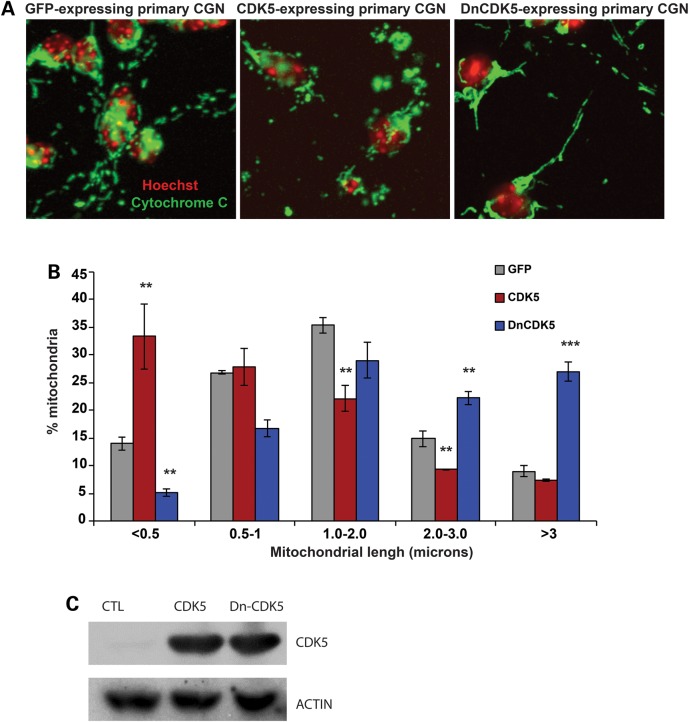
Figure 3.
Cytoplasmic CDK5 regulates mitochondrial morphology in cerebellar granule neurons. (A) Cerebellar granule neurons were infected with adenoviruses carrying expression cassettes for either of GFP-tagged cytoplasmic CDK5 (CDK5), a dominant negative cytoplasmic CDK5 (DN-CDK5), or a GFP control at the time of plating. Following 48 h in culture, neurons were fixed and mitochondrial morphology was assessed as described in Figure 2. Representative images of mitochondrial morphology in each group are shown. (B) Mitochondrial length was quantified and graphed. **P < 0.01; ***P < 0.001, Mag bar, 5 µm. (C) Protein lysates of CGN infected with GFP, CDK5-NES or Dn-CDK5-NES were analyzed by western blot analysis using antibodies to DRP1 and CDK5. Beta-ACTIN was used as loading control.

