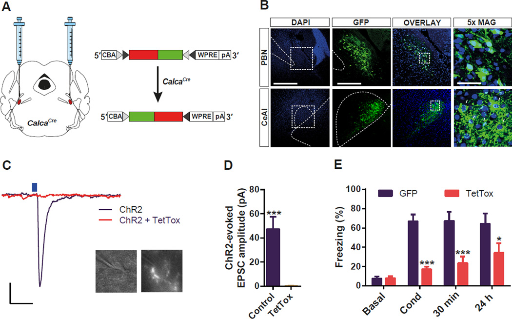
Figure 2. Functional Silencing of CGRP Neurons in the PBel Attenuates Threat Learning.
(A) Bilateral delivery of AAV carrying Cre-dependent TetTox into the PBN of CalcaCre mice.
(B) Representative histological images of TetTox expression in the CGRP neurons in the PBN (upper panels), and their terminal projections to the CeAl (lower panels). White arrows indicate their characteristic perisomatic synapses in the CeAl.
(C and D) Example traces (C) and quantification (D) of photostimulation-evoked EPSCs in the CeAl neurons that receive direct inputs from the PBel CGRP neurons. Brain slices containing the CeAl were obtained from mice previously injected with Cre-dependent ChR2 plus TetTox, or ChR2 alone into the PBN. Only neurons surrounded by fluorescent boutons were recorded (c). Scale bar: 10 pA, 25 ms. Data in (D) are means ± s.e.m. from 10 neurons (3 mice) per group.
(E) Genetic silencing of CGRP neurons in the PBel by TetTox attenuated freezing responses immediately after the conditioning (Cond), and 30 min or 24 h after the conditioning when compared with the GFPexpressing control mice. All data shown are means ± s.e.m. from 8 mice per group. *P < 0.05; **P < 0.01; ***P < 0.001.

