
Figure 3.
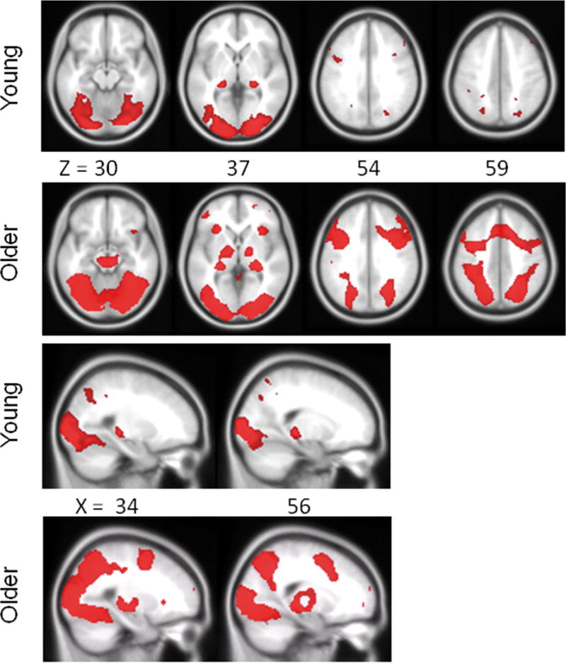
Regions of increased activation shown separately for older and young adults from the comparison Difficult Objects > Difficult Size. SPMs are thresholded at p<.05, FWE corrected for multiple comparisons. The upper two panels highlight the similar distribution of posterior ventral-visual stream regions for the two groups, as well as more extensive bilateral and medial frontal activation for older adults compared to young adults. The lower two panels show similar bilateral posterior hippocampal activation for both groups. X and Z refer to MNI coordinates for axial and saggital sections, respectively.