
Figure 4.
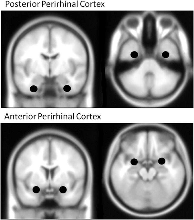
Masks for posterior and anterior PRC regions, derived from activation for the young group within the anatomical boundary of the PRC (Price and Devlin, 2007), using the contrast Difficult Objects > Difficult Size. MNI coordinates for the four regions: Posterior PRC (left −32, −4, −36; right 34, −8, −38), Anterior PRC (left −26, 4, −18; right 26, 4, −18).