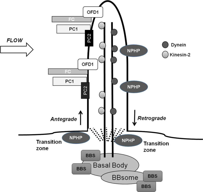
Figure 1. Localization of Human Cystic Renal Disease Proteins.

Proteins associated with ARPKD, ADPKD, Nephronophthisis, Bardet-Biedl Syndrome and Oro-facial-digital syndrome type 1 localize to primary cilia on renal epithelial cells. Proteins are transported in and out of the cilia by Kinesin-2 or IFT-dynein motors, respectively. Several NPHP proteins localize to the transition zone, the region between the cilia and the basal body. BBS -related proteins localize to the basal bodies where some aggregate to form the BBsome. Abnormal ciliary proteins are thought to interact with cilia to affect downstream signaling pathways including those related to cyclic AMP, mTOR, Canonical Wnt, G-protein coupled receptors, epidermal growth factor receptor family, MAP/ERK, planar cell polarity, calcium and cell cycle signaling. The exact mechanisms by which these interactions occur remain poorly defined.
(FC= fibrocystin; PC1 = polycystin 1; PC2 = polycystin 2; NPHP= nephrocystin; BBS = Bardet-Biedl proteins; OFD1= orofacial digital syndrome type 1)
