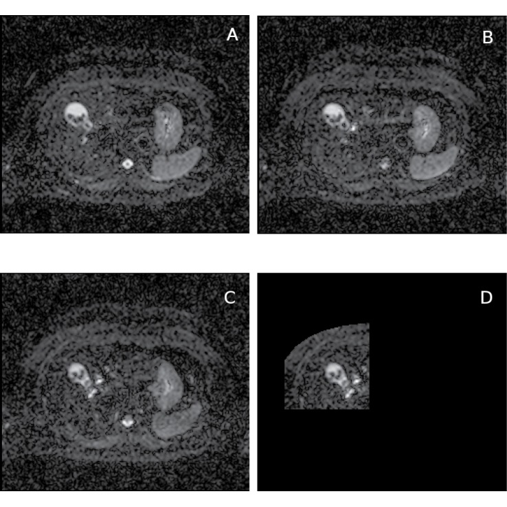
Fig 2. Diffusion gradient direction components of DW-MR data and the reference image.
Healthy liver DW-MR image data (v3-0528-slc24); b-value = 100s/mm 2; selected image slice from one of the repeat data-sets (out of 4) and diffusion gradients in y (A) and z (B) directions; diffusion gradient in −x direction which has been used as the reference image slice for local-rigid alignment (C) and the reference image shown in the selected ROI (D); this ROI is used both in local-rigid alignment and ADC histogram analysis; gamma adjusted.

