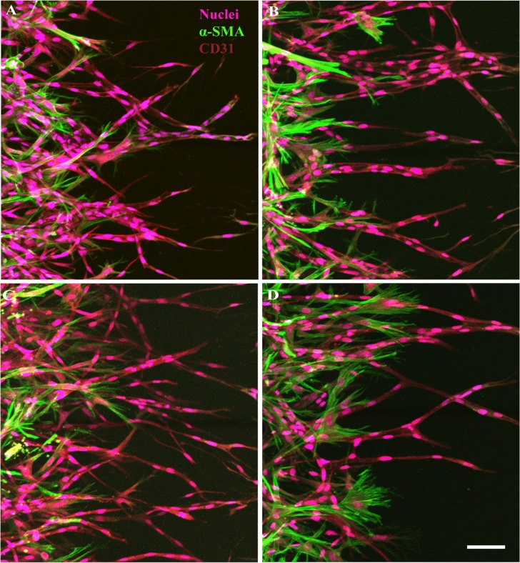
Fig 6. Effect of VEGF-A, TNFα and IL-1α on EC-pericyte association.
Representative confocal images showed contrasting morphology of the blood vessels in response to different biochemical factors. In comparison with the control condition (A), VEGF-A (100ng/ml) treated vessels were dilated and pericytes showed contracted morphology (B). Inflammatory cytokines TNFα (10 ng/ml) (C) and IL-1α (10 ng/ml) (D) treated pericytes showed both distinct filopodia growth, but in different morphology. Scale bar 50 μm.

