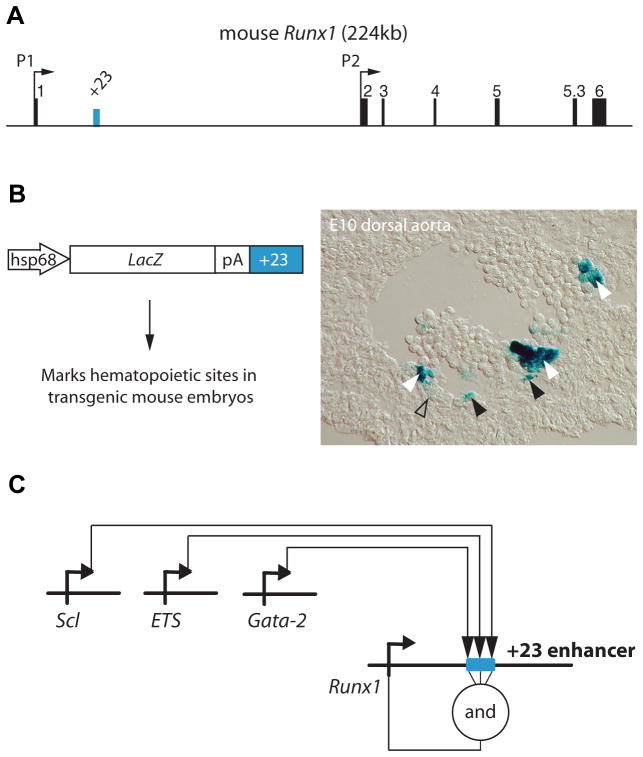
Fig. 2. The Runx1 +23 enhancer recapitulates the hematopoietic specific expression pattern of Runx1.
(A) Schematic of the Runx1 locus. Vertebrate Runx1 is transcribed from two promoters, the P1 and the P2. A 531 bp mouse-frog conserved enhancer was identified (Nottingham et al., 2007) and is located 23 kb downstream of the ATG in exon 1. (B) This +23 enhancer targets reporter gene expression to hematopoietic sites in the developing embryos, including all emerging HSCs. A transverse section through the dorsal aorta of an E10 transient transgenic embryo shows Xgal staining in emerging hematopoietic clusters (white arrowheads), in scattered cells of the endothelial wall (black arrowheads), and in a few mesenchymal cells (open arrowhead). Identical Xgal staining is seen in established mouse lines carrying the hsp68LacZ+23 transgene (not shown). (C) Targeted mutagenesis of putative transcription factor binding sites and chromatin IP (Nottingham et al., 2007), and trans-activation assays (Landry et al., 2008) placed the Runx1 +23 enhancer directly downstream of the ETS/GATA/SCL kernel (Liu et al., 2008; Pimanda et al., 2007b) that is active at the onset of developmental hematopoiesis.

