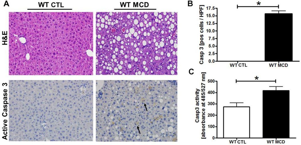
Fig. 1. MCD diet results in steatohepatitis development and marked hepatocyte caspase 3 activation.
(A) Representative pictures of Hematoxylin & Eosin (H&E) staining and active caspase 3 immunohistochemistry of liver from the wild-type animals on the MCD or control diets (magnification 40x) and corresponding quantification (B). (C) Caspase 3 activation in the different groups of mice was assessed by Apo-ONE Homogeneous Caspase 3 fluorometric assays. Results are represented as mean ± SEM. * P < 0.05.

