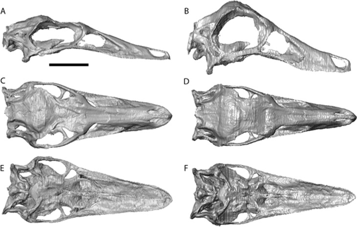
Figure 3. Struthiomimus altus reconstruction (RTMP 1990.026.0001).
Note the dorsoventral expansion of the skull after retrodeformation, particularly of the orbital region. (A), (C), (E), original skull, (B), (D), (F), retrodeformed skulls. (A),(B), right lateral; (C), (D) dorsal; (E), (F), ventral views. Scale bar = 5 cm. See Videos S3 and S4 showing video of the skull before and after retrodeformation.

