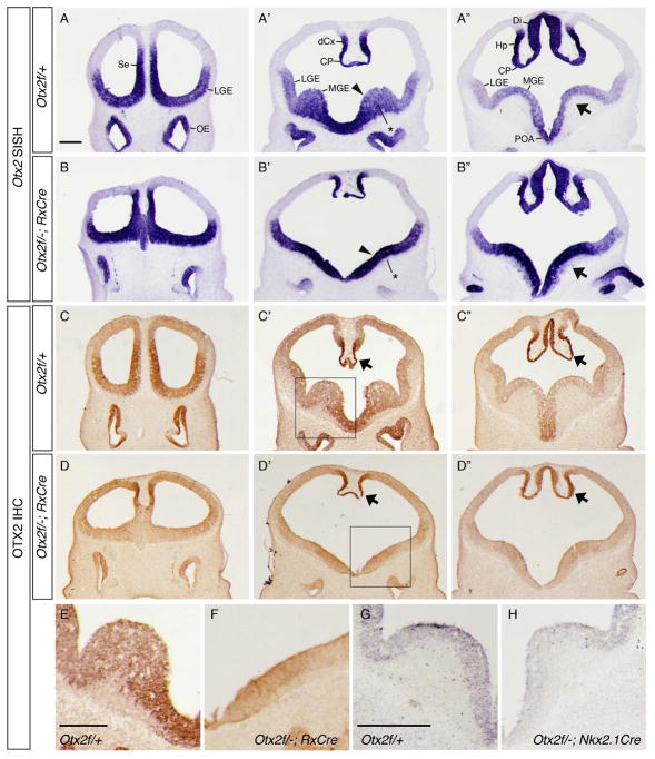
Figure 1. Otx2 expression in conditional knock outs (cKOs).
(A-B”) ISH on E11.5 coronal sections from (A–A”) Otx2f/+ and (B–B”) Otx2f/−; RxCre embryos using a full length Otx2 riboprobe. Otx2 transcription appears upregulated in the MGE of RxCre cKOs (arrowheads and arrows in A’, A”, B’, B”), and that the MGE SVZ and MZ are hypoplastic (asterisks in A’, B’). (C–H) Anti-OTX2 IHC: in RxCre cKOs (C–F), OTX2 protein expression is absent in cKO forebrains except in the dorsomedial caudal cortex, hippocampal anlage, and choroid plexus (arrows, C’-C”, D’–D”). E and F show higher magnification views of the boxed regions in C’, D’. (G–H) In Nkx2.1Cre cKOs, OTX2 expression was reduced in the MGE. A–D”: rostrocaudal series of coronal sections. Abbreviations: Se, septum; MGE, medial ganglionic eminence; LGE, lateral ganglionic eminence; dCx, dorsomedial cortex; Hp, hippocampal anlage; POA, preoptic area; Di, diencephalon; CP, choroid plexus; OE, olfactory epithelium. Scale bars: A, E = 0.25mm, G = 0.4mm.

