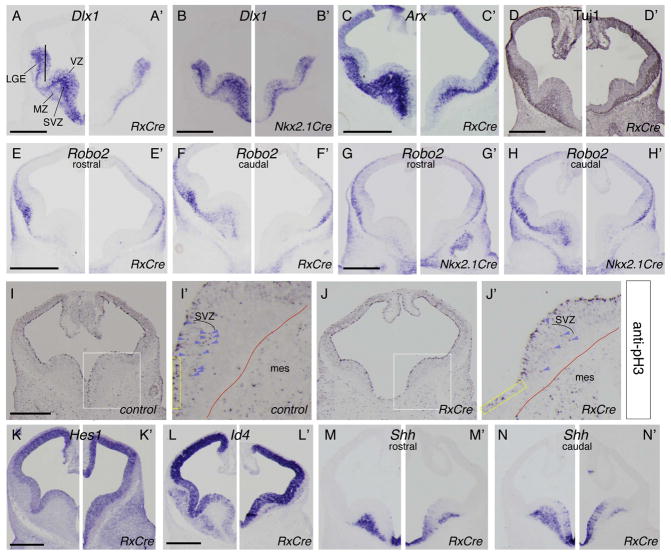
Figure 5. Reduced neurogenesis and proliferation in the basal ganglia of E11.5 Otx2 cKOs.
ISH and IHC on coronal hemisections (control: left, mutant: right). (A–H’) ISH: (A–H) Otx2f/+, (A’, C’, D’–F’) Otx2f/−; RxCre, and (B’, G’–H’) Otx2f/−; Nkx2.1Cre embryos using probes to (A–B’) Dlx1, (C–C’) Arx, (E–H’) Robo2. Anti-Tuj1 IHC (D) Otx2f/+ and (D’) Otx2f/−; RxCre. (I–J’) Anti- pH3 IHC: (I’, I) Otx2f/+ and (J, J’) Otx2f/−; RxCre embryos. I’–J’: high magnification of I, J. Red lines: neural/mesenchymal boundary; purple arrowheads: pH3+ SVZ cells; yellow rectangles highlight similar VZ regions of the vMGE in K–L’ showing upregulation of Hes1 and Id4, respectively. (K, L) Otx2f/+ and (K’, L’) Otx2f/-; RxCre embryos. (M–N’) Shh reduction in MZ and increase in VZ. Abbreviations: mes: mesenchyme; SVZ: subventricular zone; VZ ventricular zone. Scale bars: 0.5mm.

