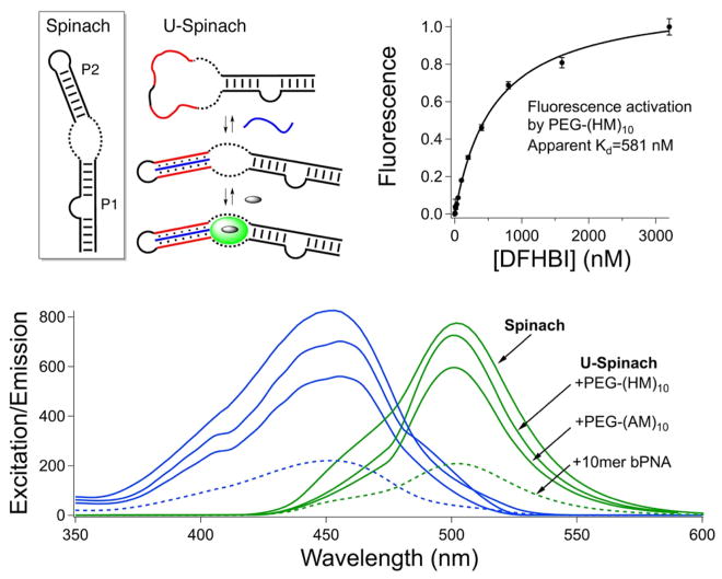
Figure 4.
Aptamer turn-on by bifacial polymer nucleic acid. (top left) Spinach aptamer fold, with the fluorogen binding site shown as a dashed line; refolding of U-Spinach by bPoNA (blue) and DFHBI binding, with U tracts shown in red. For clarity, the PEG block is not indicated. (top right) Fluorescence activation of DFHBI by the bPoNA–U-Spinach binary complex, fit to a 1:1 binding model. (bottom) Excitation (blue) and emission (green) spectra for the indicated DFHBI complexes. The excitation and emission intensities follow the same trend.

