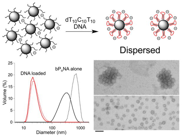
Figure 5.
(top) Aggregated bPoNA nanoparticles disperse upon binding of DNA (red). (bottom left) DLS of p(AM)10-b-PnBA nanoparticles with (red) and without (black) DNA loading after 6 h (—) and 72 h (---). (bottom right) TEM images of uranyl acetate-stained particles clustered without DNA and dispersed after DNA binding. The scale bar is 100 nm.

