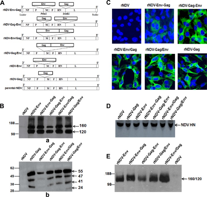
FIG 1 .
Expression analysis of gp160 and Gag in DF1 cells infected with different rNDVs. (A) Schematic representation of rNDV strain rLaSota with human codon-optimized HIV-1 p55 Gag and gp160 Env genes inserted at different positions. The restriction sites that were used to insert the Gag and the gp160 genes (PmeI and SnaB1) are indicated. Each transcriptional cassette was flanked by NDV gene start and a gene end transcription signals. (B) Expression and processing of Env and Gag proteins in DF1 cells infected with the indicated viruses. Cell lysates were analyzed 48 h postinfection by Western blotting using a pool of gp120-specific MAbs (panel a) and p24 Gag-specific MAb (panel b). (C) Immunofluorescence analysis of the intracellular expression of Gag protein in DF1 cells infected with indicated virus. The cells were probed with p24 Gag-specific MAb followed by staining with Alexa Fluor 488-conjugated goat anti-mouse IgG antibodies (green) and DAPI (4[prime],6-diamidino-2-phenylindole) (blue) and subsequently visualized under a confocal Zeiss LSM 510 fluorescence microscope. (D) Western blot analysis of NDV HN protein expression by different rNDVs in DF1 cells. (E) Analysis of incorporation of HIV-1 gp120 and 160 into purified rNDV virions using a pool of gp120-specific MAbs. The positions of the HIV gp160 precursor and its cleavage product gp120 and of the p55 Gag precursor and its cleavage products p47, p41, and p24 are indicated by arrows in the right margin in different gels.

