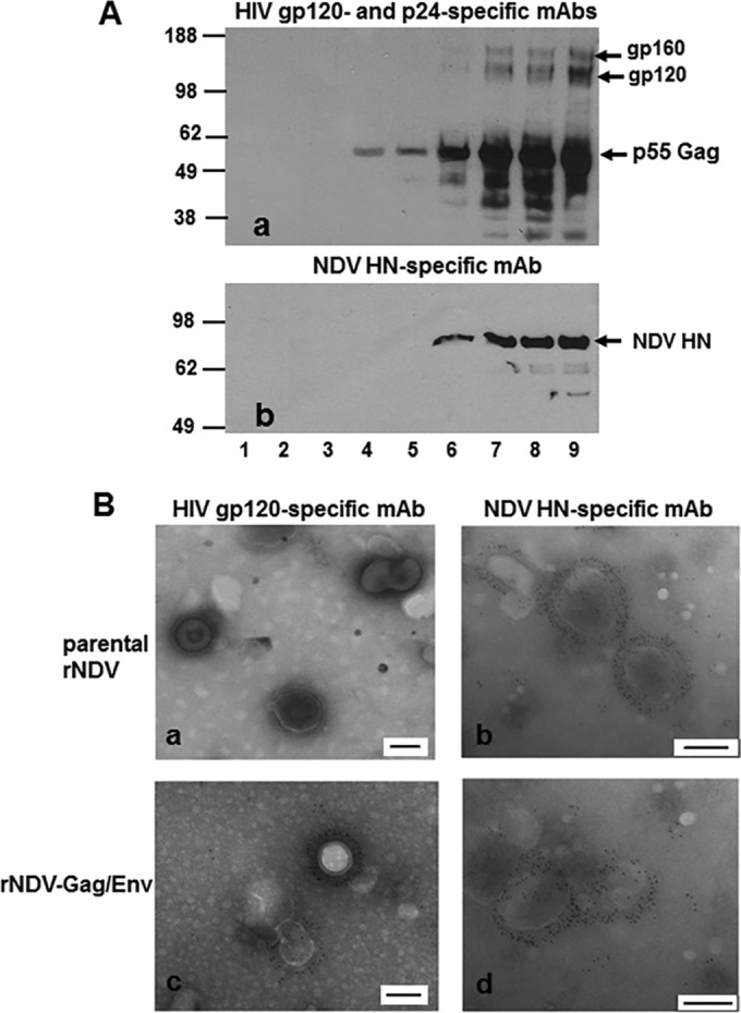
FIG 2 .

Biochemical characterization of HIV-1 VLPs produced in the allantoic fluid of 9-day-old embryonated chicken eggs infected with rNDV. (A) Western blot analysis of sucrose gradient fractions obtained after ultracentrifugation of rNDV-Gag/Env-infected allantoic fluid using HIV gp120-specific and p24-specific MAbs (panel a) or NDV HN-specific MAb (panel b). (B) Results of analysis of incorporation of gp160 (Env) protein on HIV-1 VLPs were verified by electron microscopy. Fraction 8 (shown in lane 8 in panel A) was adsorbed onto carbon-coated EM grids and stained with either anti-HIV gp120 MAb (panels a and c) or NDV HN-specific MAb (panels b and d) followed by detection with 6-nm-diameter-gold-nanoparticle-conjugated anti-mouse IgG antibodies. Each scale bar in panels a to d represents 100 nm.
