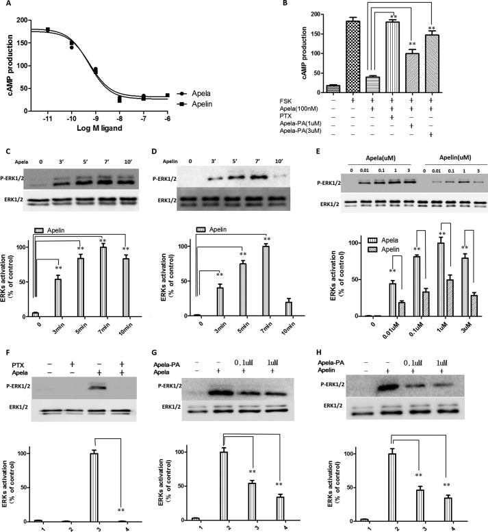
FIGURE 2.
Apela is equipotent as apelin in suppressing cAMP production but more potent than apelin in the stimulation of ERK1/2 phosphorylation mediated by the APJ receptor. A, inhibition of forskolin-stimulated cAMP production by apela or apelin in CHO cells stably expressing APJ. CHO cells expressing APJ were pretreated for 30 min with apela or apelin at indicated concentrations before treatment with 1 μm forskolin (FSK) in the presence of 0.2 mm IBMX. At 30 min later, total cAMP levels were determined. Experiments were repeated independently at least three times. Data were analyzed using Graphpad Prism 5.0 and IC50 values were calculated. IC50 of apela is 0.27 nm ±0.011, and IC50 of apelin is 0.29 nm ±0.013, respectively; Imax value of aplea is 178.8 ± 5.3 and Imax value of aplea is 183.3 ± 4.6, respectively. B, pretreatment with PTX and apela-PA abrogated the inhibition of forskolin-stimulated cAMP production by apela. To determine the effects of PTX pretreatment, cells were pretreated overnight with PTX (200 ng/ml) before testing. For determining the effects of apela-PA, cells were pretreated with apela-PA (1 μm or 3 μm) for 30 min. before testing. Experiments were repeated independently at least three times. Calculations were done with a standard statistical package (SPSS for Windows, version 21). Statistical significance was defined as p value <0.05 (*) or p value <0.01 (**). C and D, apela and apelin treatment promoted a time-dependent phosphorylation of ERKs in CHO cells expressing APJ. E, apela induced higher maximal phosphorylation of ERK1/2 than apelin. CHO cells stably expressing APJ were changed into serum-free conditions for 12 h, followed by stimulation with different concentrations of apela or apelin for 7 min or indicated time course before immunoblotting analyses (upper panel, representative blots). The density of the bands corresponding to 44 kDa and 42 kDa were quantified with an imaging densitometer. Data shown in the lower panel of C, D, and E are expressed as percentages of the maximal value and represent the mean ± S.E. of three independent experiments. Calculations were done with a standard statistical package (SPSS for Windows, version 21). Statistical significance was defined as p value <0.05 (*) and p value <0.01 (**). F, PTX pretreatment abrogated the phosphorylation of ERKs induced by apela. CHO cells stably expressing APJ were changed into serum-free conditions with or without pretreatment with PTX (200 ng/ml) for 12h before stimulation with 1 μm apela for 7 min. Cells were lysed and immunoblotting analyses were performed using specific antibodies. G and H, apela-PA dose-dependently reduced effects of both apela and apelin on ERKs phosphorylation. CHO cells stably expressing APJ were changed into serum-free conditions for 12 h and were pretreated with apela-PA (0.1 μm or 1 μm) for 2 min before stimulation with 10 nm apela or apelin for 7 min. Cells were lysed, and immunoblotting analyses were performed using specific antibodies. The density of the bands corresponding to 44 and 42 kDa was quantified with an imaging densitometer. Data shown in the lower panel of F, G, and H are expressed as percentages of the maximal value and represent the mean ± S.E. of three independent experiments. Calculations were done with a standard statistical package (SPSS for Windows, version 21). Statistical significance was defined as p value <0.05 (*) and p value <0.01 (**).

