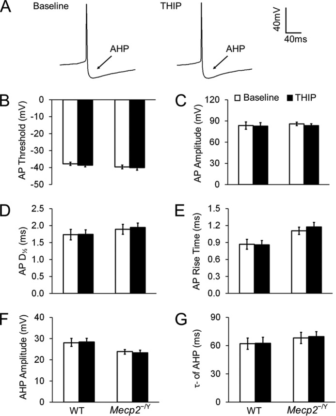
FIGURE 7.

THIP does not affect the morphology of action potential (AP) and afterhyperpolarization (AHP) in either WT or Mecp2-null neurons. A, spontaneous APs recorded from an LC neuron. No obvious changes in AP morphology were found after exposure to THIP. B–E, in the presence of ionotropic receptor blockers (AP5, 6-cyano-7-nitroquinoxaline-2,3-dione, and strychnine) in the bath solution, THIP did not change the AP threshold (the potential at the AP initiation point), AP amplitude (the amplitude from threshold to peak), rise time, and half-width (D½, measured at 50% amplitude) of APs in either WT or Mecp2-null neurons (n = 7 and 12; p > 0.05 and p > 0.05, respectively; Student's t test). F and G, in the presence of ionotropic receptor blockers, AHP was also not affected by THIP in WT and Mecp2-null neurons. AHP amplitude was measured from the AP threshold to the lowest hyperpolarization point, and the time constant of AHP was described with a single exponential in the period from 10% to 90% of the AHP amplitude (n = 7 and 12; p > 0.05 and p > 0.05, respectively; Student's t test).
