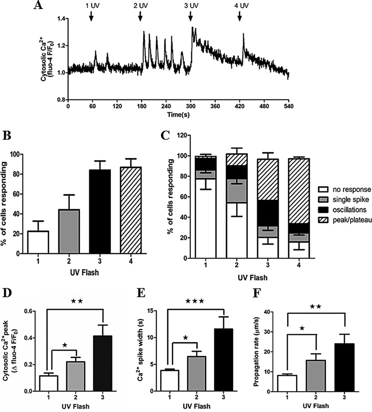
FIGURE 1.
Photorelease of caged IP3 elicits Ca2+ oscillations in primary rat hepatocytes. Isolated hepatocytes were cultured overnight and then loaded with caged IP3 and Fluo-4. A, representative trace showing cytosolic Ca2+ responses to photolysis of caged IP3. Rapid trains of one, two, three, or four UV pulses were applied as indicated (arrows). B, the percentage of cells responding to one to four UV flash events. C, comparison of the types of Ca2+ responses observed after each train of UV pulses: no response, single spike, oscillations, or a sustained Ca2+ increase (peak/plateau). Data shown are mean ± S.E. from ≥100 cells from five independent experiments. D and E, summary of Ca2+ transient amplitude (D) and Ca2+ spike width measured at half-peak height (E) in cells in which oscillations were observed after one, two, and three UV flashes. Data are mean ± S.E. of the first three Ca2+ transients for each individual cell (n = 15). F, Ca2+ wave propagation rates as a function of number of UV flashes. Data are mean ± S.E. from cells in which Ca2+ waves were observed after one, two, and three UV flashes (n = 13). *, p < 0.05; **, p < 0.01; ***, p < 0.001; Student's t test.

