FIGURE 6.
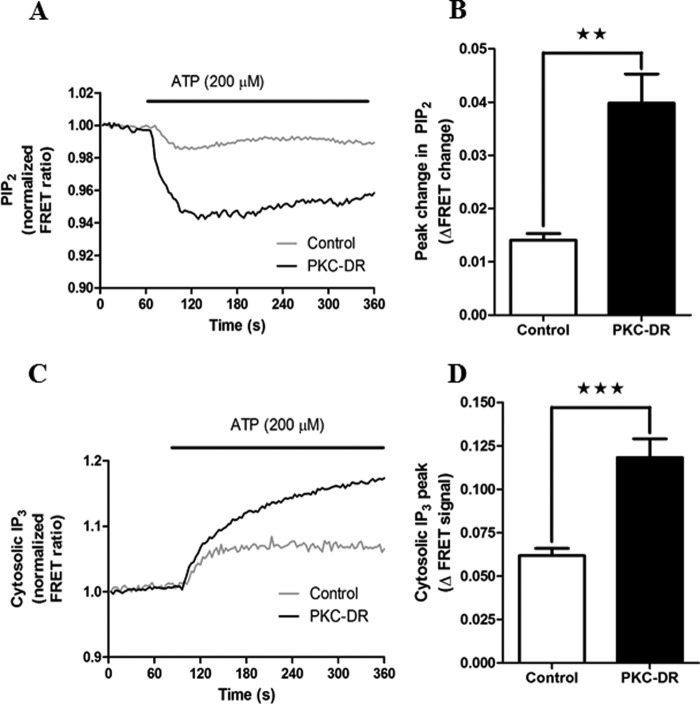
Down-regulation of PKC potentiates hormone-stimulated PLC activity and IP3 levels. Hepatocytes were transfected with eCFP-PH-PLCδ4 and eYFP-PH-PLCδ4 to monitor PIP2 levels (PLC activity) or IRIS-1 to monitor IP3 production (see “Experimental Procedures”) and then cultured overnight in the presence of 2 μm 4α-PMA (Control) or 2 μm PMA (PKC-DR). Cells were stimulated with ATP (200 μm), and maximal FRET changes were determined. A, representative traces showing the mean PLC response in control and PKC-DR cells. Traces are averaged from 27 and 25 cells, respectively, and are normalized to the basal FRET level (mean basal FRET values were 0.85 ± 0.011 for control and 1.028 ± 0.066 for PKC-DR cells, p = 0.033, Student's t test). B, mean peak FRET change (absolute values) ± S.E. for ≥70 cells from five independent experiments. **, p < 0.01, Student's t test. C, representative experiment showing the effect of PKC down-regulation on ATP-induced increases in IP3 levels for control and PKC-DR cells. Traces are averaged from five and four cells, respectively, and are normalized to the basal FRET level (mean basal FRET values were 0.66 ± 0.016 for control and 0.70 ± 0.013 for PKC-DR cells, p = 0.053, Student's t test). D, mean peak FRET change (absolute values) ± S.E. for ≥25 cells from 10 independent experiments. ***, p < 0.001, Student's t test.

