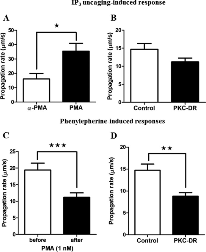
FIGURE 9.

Ca2+ wave velocity is regulated by PKC activity. Isolated hepatocytes were treated overnight with 4α-PMA (1 μm, Control) or PMA (1 μm, PKC-DR) or treated acutely with 1 nm PMA to assess the effect of PKC on Ca2+ waves initiated by phenylephrine or caged IP3. Ca2+ wave propagation rates were calculated in micrometers per second by determining time at half-peak height from regions of interest from the wave initiation site and the opposite pole of the hepatocyte. A and B, the effect of acute PKC activation (1 μm PMA, A) and PKC-DR down-regulation (B) on caged IP3 (three UV flashes) induced Ca2+ wave velocity (micrometers per second ± S.E. for ≥16 cells from three independent experiments). C and D, the effect of acute PKC activation (1 nm) (C) and PKC-DR (D) on Ca2+ wave propagation rate in response to phenylephrine. Data are mean wave velocity (micrometers per second) ± S.E. for ≥18 cells from four independent experiments and from ≥20 cells from three independent experiments, respectively. *, p < 0.05; **, p < 0.01; ***, p < 0.001; Student's t test.
