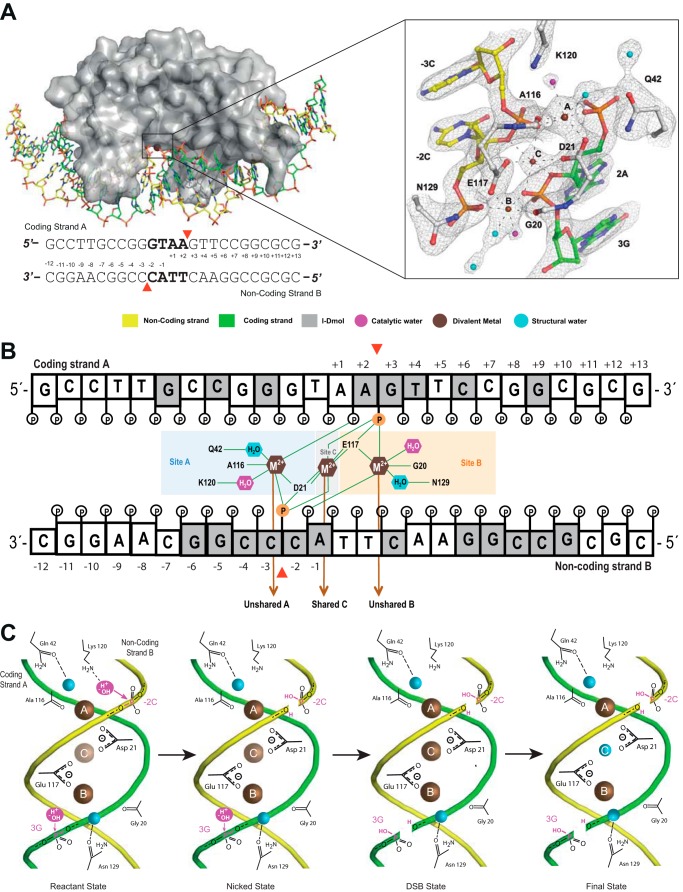
FIGURE 1.
I-DmoI active site. A, detailed scheme of the I-DmoI active site at the reactant state (Protein Data Bank code 4UN9) including the 2Fo − Fc map contoured at 1.2σ. The sequence of the DNA target is shown below. Red arrowheads indicate the cleavage sites. B, scheme of the active center contacts. Bases are colored in gray or white depending on whether they contact the protein amino acids or not, respectively. The red arrowheads indicate the positions of the targeted phosphodiester bonds (orange). Catalytic waters are shown in magenta. C, schematic diagram summarizing the catalytic mechanism. The color key is the same as in A. Metal ion occupancy at site C is depicted by the color intensity (brown), revealing its crucial role in catalysis, triggering the consecutive hydrolysis of the targeted phosphodiester bonds in DNA strands and leaving its position once a double strand break is generated.

