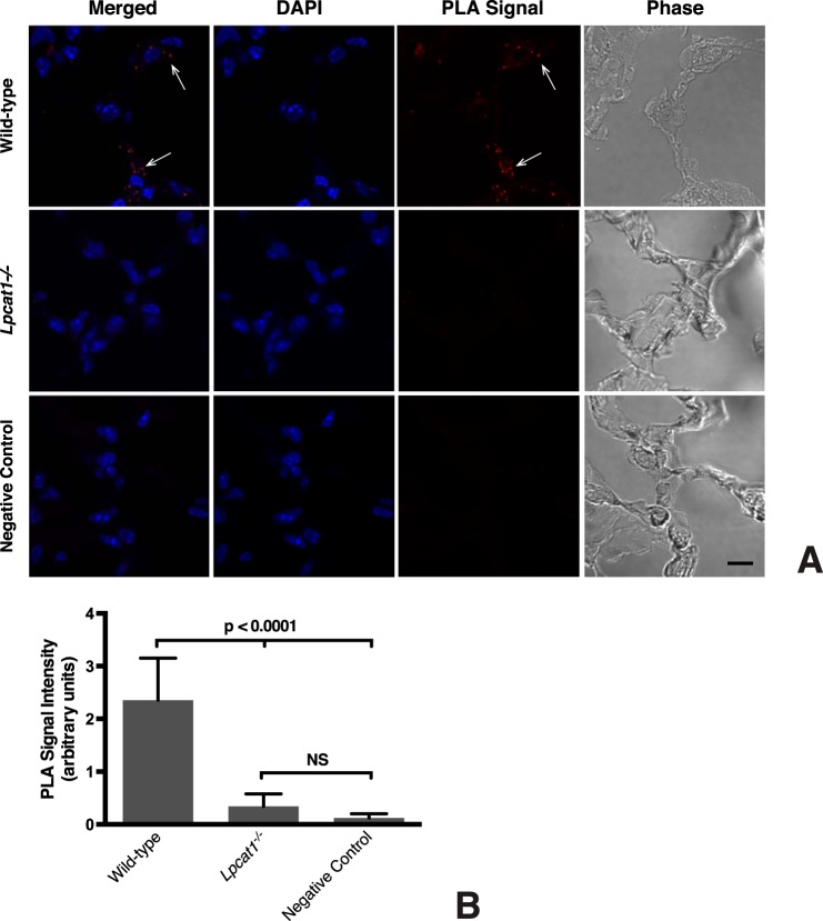
FIGURE 3.
LPCAT1 interacts with StarD10 in vivo. A, cryostat sections of frozen fixed adult lungs were subjected to in situ PLA using antibodies against LPCAT1 and StarD10. Confocal images of the PLA signal (red dots; arrows) shows that LPCAT1 and StarD10 interact in wild-type mouse lung (top row). No signal is apparent in sections of lungs from Lpcat1−/− mice (middle row) or in sections in which normal rabbit IgG was substituted for anti-LPCAT1 (negative control; bottom row). The images are representative of lung sections from two different mice for each group in five independent experiments. Scale bar, 10 μm. B, quantification of PLA signals. The values represent the mean ± S.E. (error bars) of five independent experiments in each group. p < 0.0001 versus Lpcat1−/− lungs and the negative control. NS, not significant (p ≥ 0.05).

