FIGURE 6.
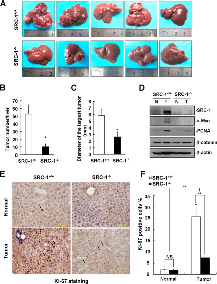
SRC-1−/− mice were resistant to DEN/CCl4-induced liver tumor formation. A, images of livers with surface tumors from SRC-1+/+ and SRC-1−/− mice after 25 weeks of DEN/CCl4 treatment. B, quantitation of the numbers of surface liver tumors from SRC-1+/+ and SRC-1−/− mice (*, p < 0.05; n = 5). C, quantitation of the diameter of the largest surface liver tumors from SRC-1+/+ and SRC-1−/− mice (*, p < 0.05; n = 5). D, c-Myc and PCNA protein levels were significantly decreased in liver tumors from SRC-1−/− mice compared with SRC-1+/+ mice. Liver tissue lysates from five SRC-1+/+ or SRC-1−/− mice were mixed together for Western blot analysis of SRC-1, c-Myc, PCNA, and β-catenin. β-Actin was used as a loading control. N, non-tumorous tissue; T, tumor tissue. This experiment was performed three times with similar results. E, Ki-67 staining of tissue sections from non-tumorous liver tissues and liver tumors from SRC-1+/+ and SRC-1−/− mice. A representative image is shown. F, quantitation of Ki-67-positive tumor cells (**, p < 0.01; NS, not significant; n = 4 tumors/group). Error bars, S.E.

