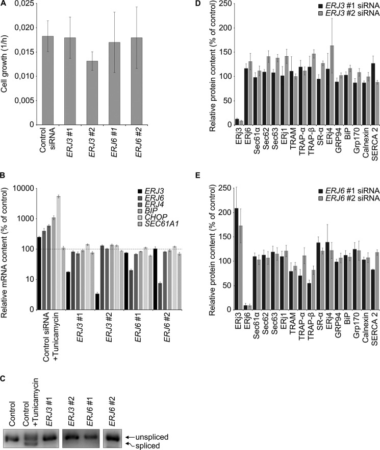
FIGURE 3.
Effect of ERJ3 and ERJ6 gene silencing on cell proliferation and the content of selected mRNAs and proteins. A, HeLa cells were treated with the indicated siRNAs for 48 h and seeded in E-plates. Proliferation was recorded in real time for 48 h. Growth rates were measured in three independent experiments in triplicate and are given with S.E. (error bars) as slope of the curves for the second 48 h. Silencing was evaluated by Western blots (data not shown). B–E, HeLa cells were treated with the indicated siRNAs for 96 h as in Fig. 2, and their content of selected mRNAs and proteins, respectively, was evaluated by quantitative real-time PCR (B), agarose gel electrophoresis (C), or Western blots (D and E). B and C, as positive control for UPR activation, control siRNA-treated cells were treated with tunicamycin for 5 h at 2 μg/ml. B, averaged relative mRNA contents from four independent experiments are given with S.E. as a percentage of control siRNA-treated cells and as normalized to ACTB. The 100% values are indicated by a dashed line. C, XBP1 was amplified in the same cDNA as in B with appropriate primers and subjected to gel electrophoresis and imaging. Only the area of interest from a single 3% agarose gel is shown. Lanes 4 and 5 represent a longer exposure as compared with the other lanes. D and E, for Western blots, averaged relative protein contents are given with S.E. as a percentage of control siRNA-treated cells and as normalized to β-actin.

