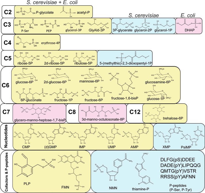
FIGURE 10.
Substrate diversity of S. cerevisiae and E. coli HADs; the molecular structures of positive HAD substrates. Light yellow background, positive substrates common for both organisms; light blue background, substrates shown to be positive only for S. cerevisiae HADs; light pink background, substrates shown to be positive only for E. coli HADs.

