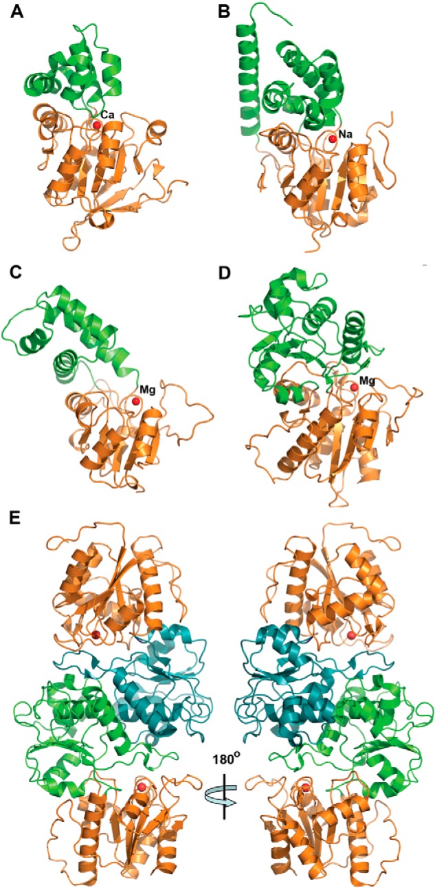
FIGURE 5.

Crystal structures of four S. cerevisiae HADs. Overall structures of the protomers: RHR2 (A), SDT1 (B), UTR4 (C), and YKR070W (D). All enzymes show a two-domain organization with the Rossmann-like protein core (orange ribbons) and mostly α-helical cap domain (green ribbons). In all structures, the position of potential active site is indicated by the bound metal ion (shown as a red sphere and labeled). E, two protomers of YKR070W form a tight dimer through the interaction of their cap domains (colored green and cyan, two views related by a 180° rotation).
