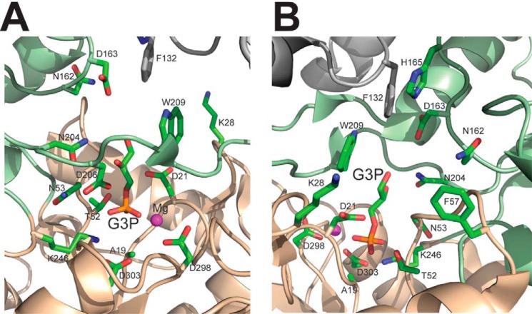
FIGURE 9.
Close-up view of the active site of the YKR070W dimer with the bound Gly-3-P (A and B, two views related by a 180° rotation). The core and cap domains of one protomer are colored in orange and light green, respectively, with amino acid side chains shown as sticks along a protein ribbon, whereas the ribbon and Phe-132 of the second protomer are colored in gray. The side chain of Phe-132 of one protomer contributes to the substrate binding site of another protomer.

