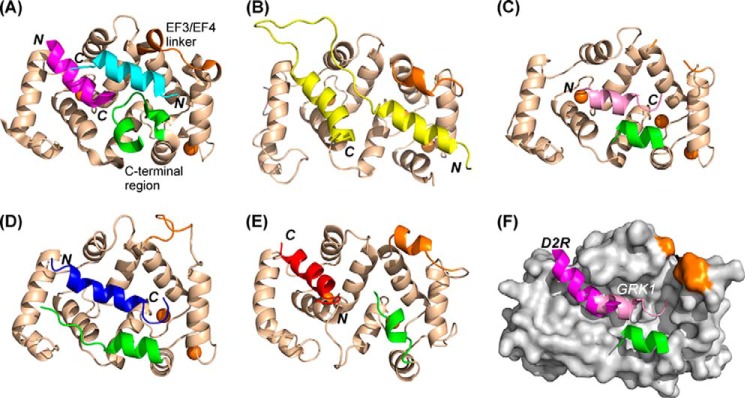
FIGURE 10.

Comparison of the structures of NCS protein complexes. Schematic representations are shown of the structures of NCS-1 in complex with two molecules of D2R peptide (magenta and cyan) (PDB 5AER) (A), ScNcs1in complex with fragment of Pik1 (yellow) (PDB 2JU0) (45) (B), NCS-1 in complex with one molecule of GRK1 peptide (pink) (PDB 5AFP) (C), and KChIP1 with a fragment of bound Kv4.3 (blue) (PDB 2I2R) (50) (D). E, recoverin bound to the N terminus of GRK1 residues 1–25 with GRK1 peptide (red; PDB 2I94) (52). F, overlay of structures 5AER and 5AFP showing the locations of the D2R bound in the N-site and GRK1 peptides. The peptide orientations are indicated as N and C in bold italics, and the orientations of the NCS protein are identical in all the structures. The EF3/EF4 linker is colored brown and the C-terminal region green; for clarity these regions are indicated only for the NCS-1·D2R peptide complex. In all the structures, Ca2+ ions are shown as brown spheres.
