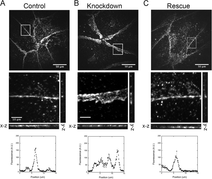
FIGURE 4.
Top row, depletion of N-WASP increases the apparent width of cell-cell junctions in x-y projection images in the upper row of images, based on anti-VE-cadherin staining. Middle row, orthogonal views of x-z and y-z image planes. Bottom row, representative line scans from the vertical (y-z) lines. A, control endothelial monolayers have thin bands of VE-cadherin at cell-cell junctions. Scale bar, 50 μm. B, N-WASP-depleted monolayers show thick bands of VE-cadherin at junctions in the x-y plane image. However, the band is thin in the x-z and y-z image planes. Scale bar, 10 μm. C, expression of full-length N-WASP restores VE-cadherin junctional bandwidth to that of control monolayers.

