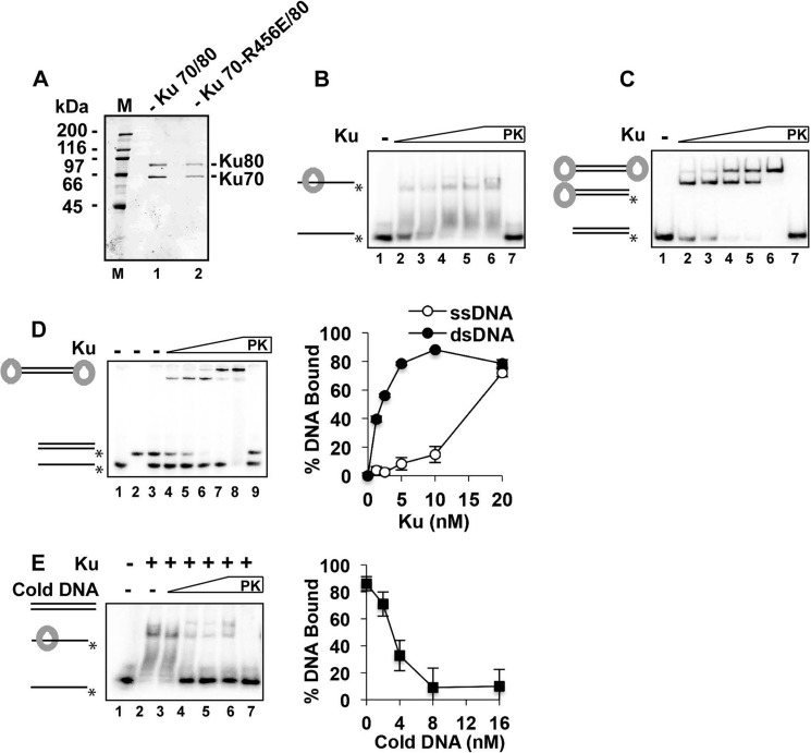
FIGURE 1.
Binding of ssDNA by Ku. A, SDS-PAGE analysis of purified Ku and the Ku-RE mutant. Molecular masses are indicated in kilodaltons (M lane). B, Ku (1.25, 2.5, 5, 10, and 20 nm) was incubated with the 40-mer ssDNA (4 nm) for 10 min. C, same conditions, except that the 40-bp dsDNA was tested. D, same conditions, except that both the 40-mer ssDNA and 40-bp dsDNA substrates were included in the same reactions. Quantification of the results is shown to the right. E, Ku (5 nm) was incubated with the radiolabeled 40-mer ssDNA (4 nm) for 10 min, and unlabeled 40-bp dsDNA (0, 2, 4, 8, and 16 nm) was added, followed by a further 10 min of incubation. Quantification of the results is shown to the right. In B–E, PK lanes represent a reaction that was treated with SDS and proteinase K prior to analysis, and asterisks denote the 32P label. Error bars in D and E represent 1 S.D. based on results from three independent experiments.

