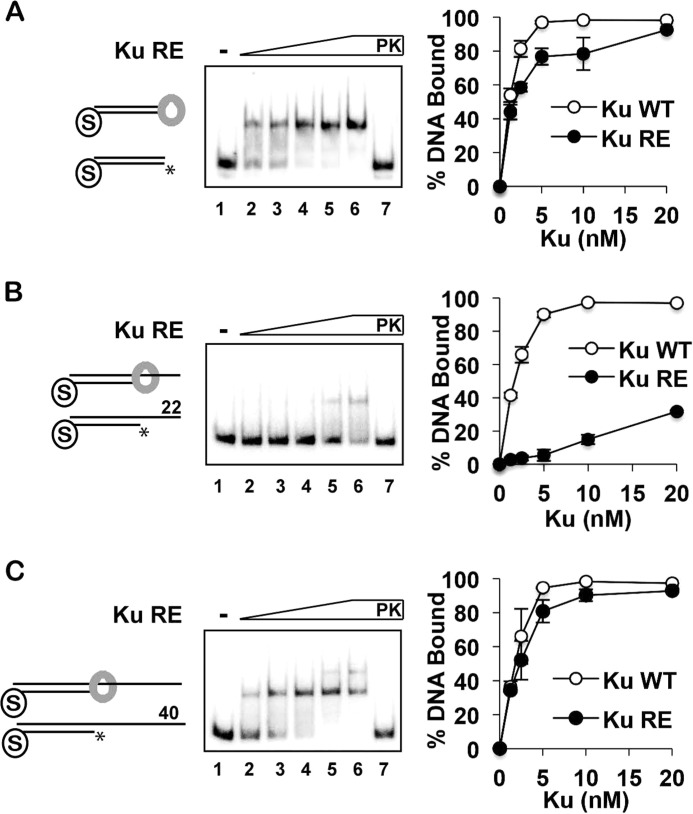
FIGURE 5.
Examination of the Ku-RE mutant for DNA end binding. A–C, Ku-RE (1.25, 2.5, 5, 10, and 20 nm) was incubated with DNA substrates (4 nm each) that had a blocked end and also a free blunt end (A) or a 22-nt (B) or 40-nt (C) 5′-recessed end. The quantification compares the results from these experiments with those obtained with wild-type Ku (from Fig. 2). PK lanes represent a reaction that had been treated with SDS and proteinase K prior to analysis, and asterisks denote the 32P label. Error bars represent 1 S.D. based on results from three independent experiments.

