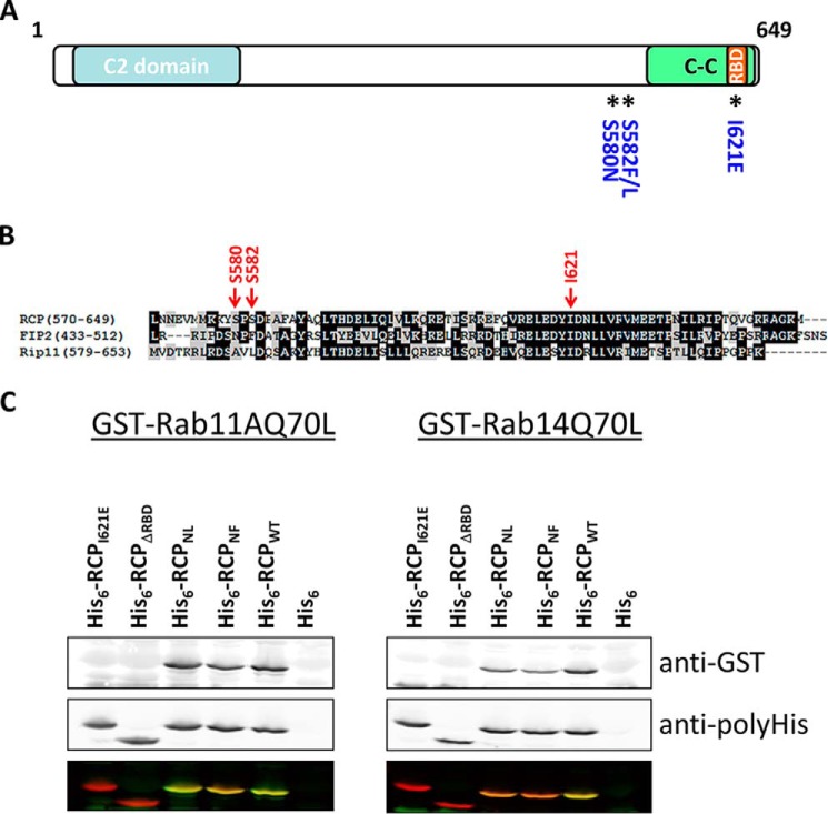
FIGURE 5.
Rab14 interacts with the RBD of RCP. A, schematic of RCP indicating conserved domains and the location of mutated residues. B, ClustalW alignment of the C-terminal region of the class I FIPs. C, far Western protein-protein interaction assays. Equal amounts of His6-fused RCP(385–649) and mutants were separated by SDS-PAGE, transferred to nitrocellulose, and overlaid with GTPγS-loaded GST-Rab11AQ70L (left) and GST-Rab14(Q70L) (right). Top panel shows the bound Rabs, revealed with an anti-GST antibody. Middle panel demonstrates equal loading of the His6-RCP wild-type and mutant fusion proteins, revealed with an anti-His antibody. Lower panel is an overlay of the anti-GST (green) and anti-His (red) immunoblots.

