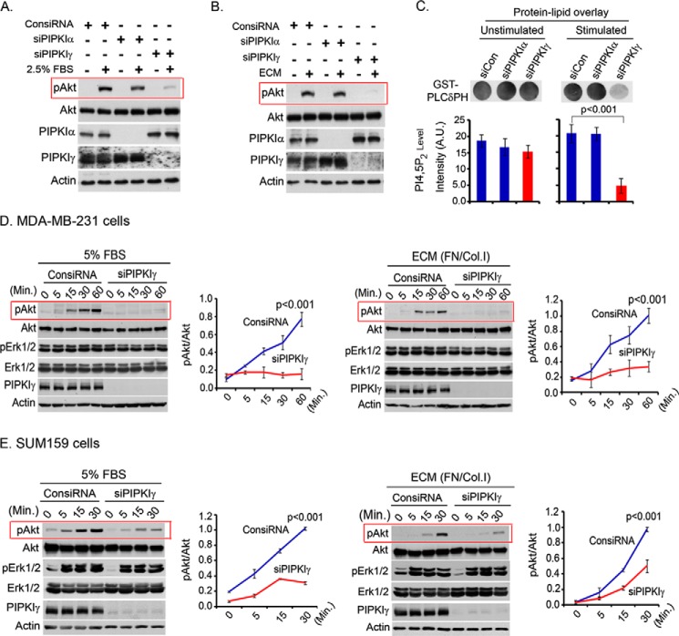
FIGURE 1.
PIPKIγ knockdown blocked Akt activation upon FBS or ECM stimulation. A and B, MDA-MB-231 cells were transfected with isoform-specific siRNA for PIPKI knockdown. 48 h post-transfection, cells were serum-starved overnight and resuspended into the serum-free medium. Cells were stimulated with FBS (A) or ECM protein (B) for 10 min in the suspension condition. Activated Akt was examined by immunoblotting using phospho-specific antibody for activated Akt. C, PIP2 level in the cells was examined by a protein-lipid overlay assay. Acidic lipids were isolated from an equal number of cells as described under “Experimental Procedures.” The isolated lipids were spotted into the nitrocellulose membrane before incubating with purified GST-PLCδPH protein. HRP-labeled anti-GST antibody was used to detect the bound GST-PLCδPH. A.U., arbitrary units. D, siRNA was used to knock down PIPKIγ expression in MDA-MB-231 cells. As described above, cells were stimulated with FBS or ECM for different time periods before examining the activated Akt and Erk1/2 by immunoblotting. The same experiments were repeated using SUM159 cells (E). Results are represented as means ± S.D. (error bars) from three independent experiments.

