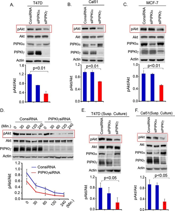
FIGURE 2.
Persistence of PI3K/Akt signaling is impaired upon the loss of PIPKIγ. A–C, isoform-specific siRNA were used to knock down PIPKI expression in T47D, Cal51, and MCF-7 cells. Cells were harvested 48–72 post-transfection to examine the activated Akt level by immunoblotting. D, T47D cells after siRNA transfection for PIPKIγ knockdown in the adherent condition were incubated in serum free-medium for different time periods before examining the activation level of Akt by immunoblotting. E and F, isoform-specific siRNA was used to knockdown PIPKI expression in T47D and Cal51 cells. 48 h post-transfection, cells were resuspended and cultured in the suspension condition by seeding the cells into culture plates precoated with soft agar. Cells were harvested 1–2 days later to examine the activation level of Akt in suspension culture. Results are represented as mean ± S.D. (error bars) from three independent experiments.

