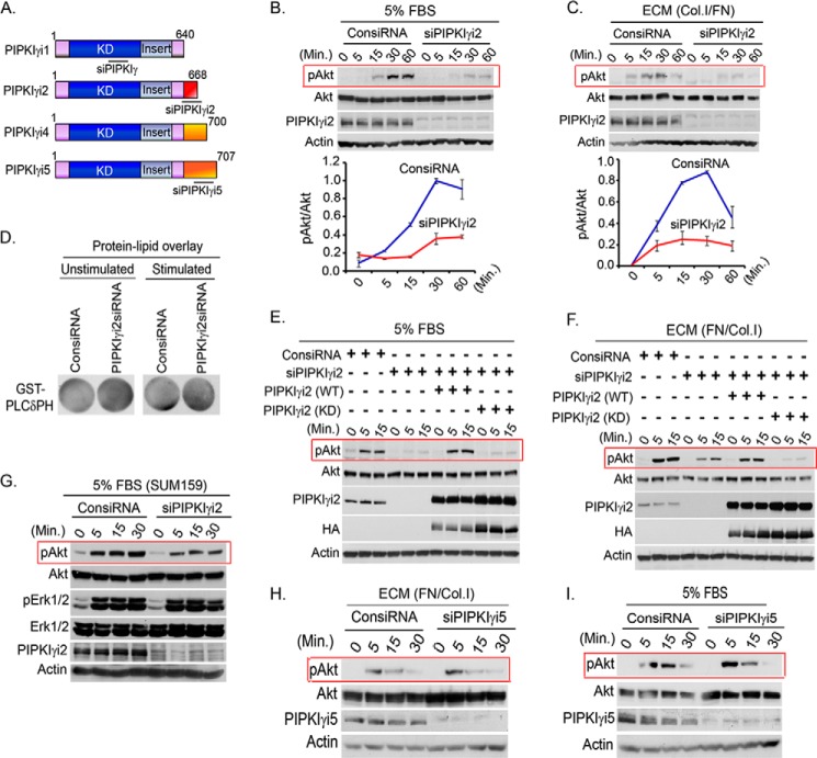
FIGURE 3.
PIPKIγi2 knockdown impaired PI3K/Akt signaling. A, schematic diagram of PIPKIγ variants and target sites for the siRNA used are indicated. B and C, MDA-MB-231 cells were transfected with siRNA specific for PIPKIγi2. 48–72 h post-transfection, cells were serum-starved overnight and resuspended into serum-free medium before stimulating with FBS or ECM for different time periods. The activation level of Akt was examined by immunoblotting using phospho-specific antibody as described above. D, PIP2 level in the cells was examined by protein-lipid overlay assay as described above. E and F, endogenous PIPKIγi2 was silenced using siRNA from MDA-MB-231 cells or MDA-MB-231 cells overexpressing siRNA-resistant PIPKIγi2 (WT) or its kinase dead mutant, PIPKIγi2 (KD). Cells were stimulated with FBS and ECM protein as described above before examining the activated Akt. G, siRNA was used to knockdown PIPKIγi2 in SUM159 cells before examining its effect on the activation level of Akt and Erk1/2 upon stimulation with FBS as described above. H and I, MDA-MB-231 cells transfected with siRNA specific for PIPKIγi5 were stimulated with FBS and ECM protein as described above, and the activation level of Akt was examined as described above.

