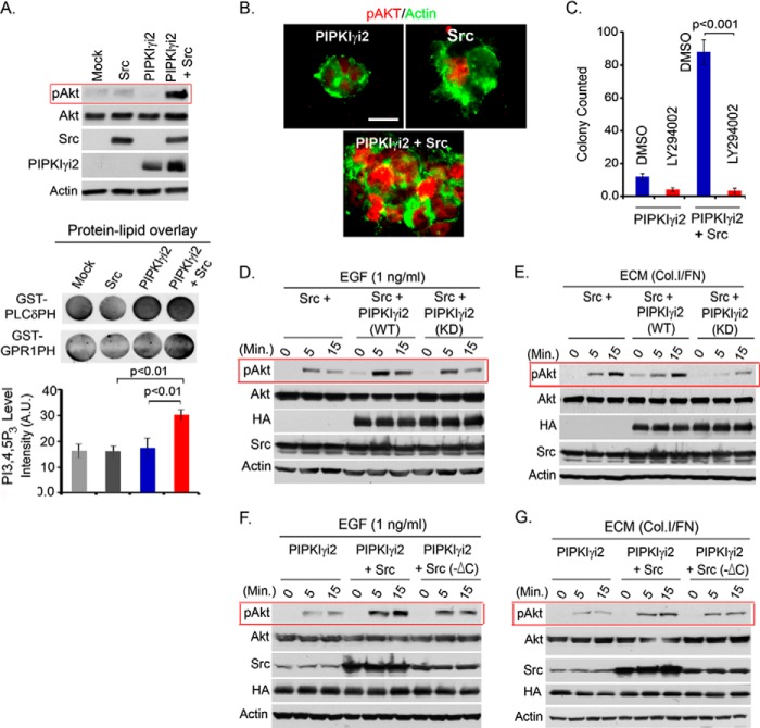
FIGURE 7.
Co-expression of PIPKIγi2 and Src induces Akt activation and oncogenic growth. A, PIPKIγi2 and Src were individually expressed or co-expressed into MDA-MB-231 cells. These cells were resuspended and cultured in the suspension condition for 1–2 days before harvesting the cells. Activated Akt was examined by immunoblotting. Similarly, lipids were extracted from these cells after 4–5 h of incubation in the suspension condition as described above. Lipids were spotted into the nitrocellulose membrane before incubating with purified GST-PLCδ-PH or GST-GRP1-PH protein. HRP-labeled anti-GST antibody was used to detect the proteins bound to lipids in the membrane. A.U., arbitrary units. B, colony developed by MDA-MB-231 cells individually expressing PIPKIγi2 and Src or co-expressing both of them was immunostained using antibody for activated Akt (red) and actin (green). Bar, 10 μm. C, anchorage-independent growth of MDA-MB-231 cells co-expressing PIPKIγi2 and Src was examined in the presence or absence of the PI3K inhibitor LY294002. Colonies formed were counted after 2 weeks of culture in soft agar as described previously. D and E, MDA-MB-231 cells expressing Src or co-expressing Src with PIPKIγi2 or its kinase-dead mutant were stimulated with EGF and ECM protein in the suspension condition before examining the activated Akt by immunoblotting. F and G, MDA-MB-231 cells expressing PIPKIγi2 or co-expressing PIPKIγi2 with wild type Src or C-terminal deletion mutant, Src (-ΔC) deficient in PIPKIγi2 binding were stimulated with EGF and ECM protein in the suspension condition before examining the activated Akt.

