Figure 3.
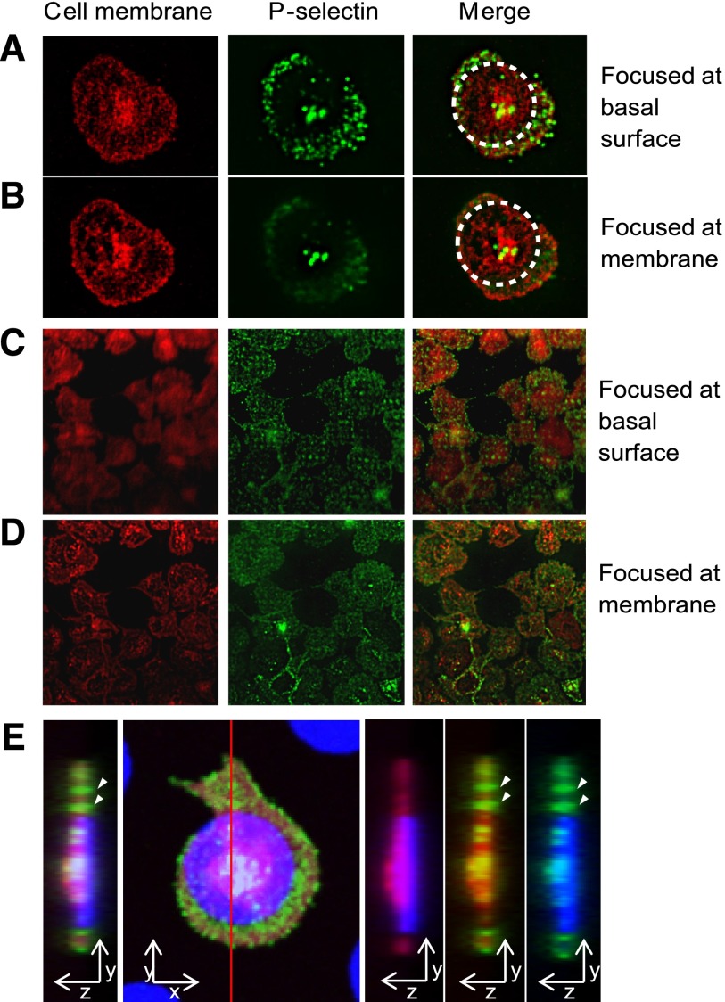
Spatially regulated α-granule secretion occurs at the basal platelet surface. (A) Total internal reflection fluorescence (TIRF) microscopy reveals that on platelets adhered and spread onto fibrinogen microdots (traced with dotted circle), anti–P-selectin staining (green) was clearly focused at the basal surface of the platelet membrane (red, cell membrane stain). (B) As the TIRF focal plane is elevated, the focus of the outer edges of the platelet membrane and anti–P-selectin staining at the granulomere center of the platelet sharpens, whereas the anti–P-selectin staining beyond the micropattern boundaries loses focus. (C) P-selectin expression localized to the submicron “holes” of the micropatterned protein matrix is clearly focused at the basal surface of the platelet membrane, whereas this focus is lost as the focal plane is elevated and focused at the outer edges of the platelet membrane (D). (E) The orthogonal sectional view of a platelet (red) on a fibrinogen microdot (blue). Strong expression of P-selectin (green) is detected in areas where platelet spreading extended beyond the geometric constraints of the microdot pattern, as well as at the granulomere center of the platelet. P-selectin expression beyond microdot pattern was also observed at the glass surface (arrows). All samples are permeabilized upon staining.

