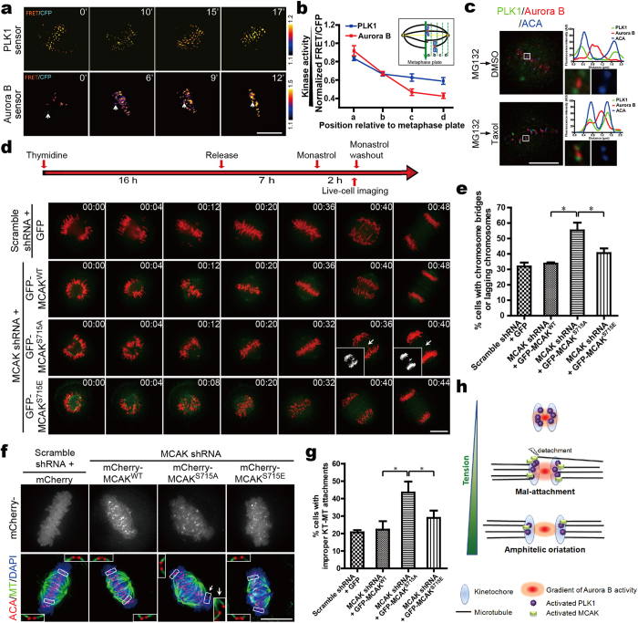
Figure 6. Precise regulation of Aurora B-PLK1-MCAK axis ensures proper kinetochore bi-orientation.
(a) Color-coded images of HeLa cells expressing PLK1 or Aurora B sensor show chromosome alignment after Syntelin washout. Cells were treated with 1 μM Syntelin for 30 min followed by three washes for subsequent real-time imaging analyses. Timestamps are in minutes. (b) Quantitative analysis of FRET/CFP emission ratio on centromeres relative to the metaphase plate. Schematic illustration of centromere position calculation (see also Supplementary Fig. S5a). (c) HeLa cells were treated with MG132 for 1 hr to allow for metaphase plate formation. Parallel samples were then treated with 1 μM Taxol or DMSO for 40 min before staining for PLK1 (green), Aurora B (red) and ACA (blue). Insets show individual kinetochore pairs used for line scans. (d) Live-cell imaging of chromosome segregation in GFP-MCAK constructs-addback cells. Cells were treated according to the protocol outlined in the upper panel. Arrows indicate lagging chromosomes during segregation. Timestamps in hr:min. (e) Quantitative analysis of the defective anaphases seen in (d). Data are presented as means ± SD from three independent experiments. *P < 0.05, Student’s t-test. (f) Cold-stable KT-MT attachment in mCherry-MCAK mutants-addback cells. Cells treated with Monastrol were released into MG132-containing medium for another 1 hr, and then fixed and stained for MTs (green), ACA (red) and DAPI (blue). Arrows indicate erroneous kinetochore attachments. (g) Quantitative analysis of cells exhibiting one or more aberrant attachments in various mutant MCAK-expressing cells. Data are presented as means ± SD from three independent experiments. *P < 0.05, Student’s t-test. (h) Model of accurate regulation of MCAK by Aurora B-PLK1 axis at kinetochore. Aurora B locates closely to PLK1 at prophase, enabling PLK1 to be activated by Aurora B at inner-centromeres (upper row). During the prometaphase-to-metaphase transition, stretch on kinetochore pairs may separate PLK1 and MCAK from Aurora B to outer-kinetochores. The stimulated MCAK activity ensures error correction in KT-MT attachment (middle row). Once the amphitelic attachments are generated, both activities of PLK1 and MCAK are down-regulated, and anaphase begins with chromosomes segregating towards the opposite poles (lower row). Scale bars, 10 μm (all image panels).

