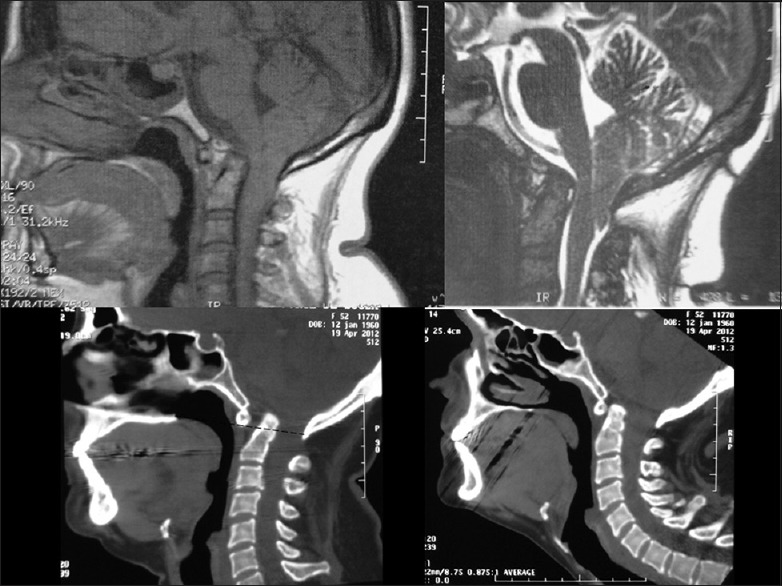
Figure 10.

Upper part: T1 (left) and T2 acquisitions showing BI1. Note the ventral cord and brainstem compression by odontoid process. Lower part: CT scan sagittal reconstruction. At left, the odontoid process in inside foramen magnum (Dotted line passing below anterior assimilated C1 arc)
