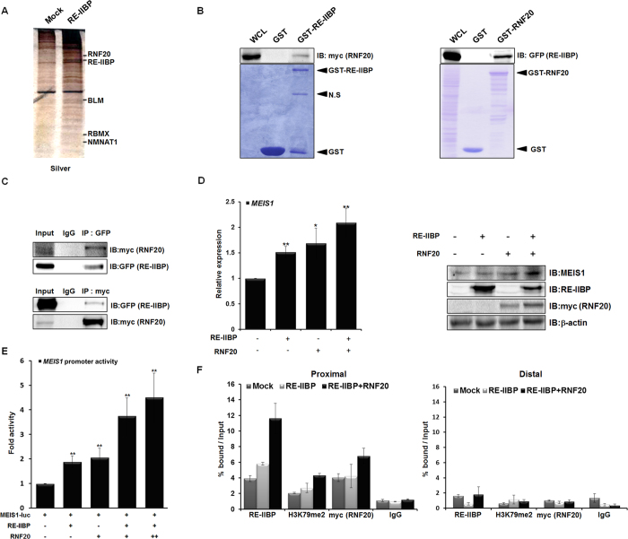
Figure 3. RNF20 Further Enhances Transcriptional Activation of MEIS1 by RE-IIBP.
(a) Binding proteins to RE-IIBP were precipitated from cell extracts of 293T cells transfected with RE-IIBP. Differentially bound proteins were detected with silver staining and identified by LC-MS analysis. (b) Extracts from 293T cells transfected with myc-RNF20 or GFP-RE-IIBP were incubated with purified GST, GST-RE-IIBP or GST-RNF20. Associated proteins were eluted, resolved by SDS-PAGE, and immunoblotted by anti-myc and anti-GFP antibodies. The amount of RE-IIBP (left) and RNF20 (right) was determined by Coomassie staining. (c) Extracts from 293T cells transfected with GFP-RE-IIBP or myc-RNF20 were immunoprecipitated with anti-GFP, and anti-myc antibodies. Associated proteins were eluted, resolved by SDS-PAGE, and immunoblotted with anti-myc and anti-GFP antibodies. (d) MEIS1 mRNA levels were analyzed using real-time PCR in RE-IIBP and RNF20 transfected 293T cells (left). Cells were lysed and immunoblotted with anti-MEIS1, anti-RE-IIBP and anti-myc antibodies (right). β-actin was used as a loading control. Results are shown as means ± SDs, n = 3; *p < 0.05, **p < 0.01. (e) 293T cells were transfected with pGL3-MEIS1 promoter reporter and the indicated DNA constructs. Cell extracts were then assayed for luciferase activity. Luciferase activity was normalized to that of β-galactosidase. Results are shown as means ± SDs, n = 3; **p < 0.01. (f) DOT1L stably knocked-down 293T cells were transiently transfected with RE-IIBP and myc-RNF20. Following transfection, ChIP analysis was performed employing anti-IgG, anti-RE-IIBP, anti-H3K79me2, and anti-myc antibodies. The immunoprecipitated DNA fragments were analyzed by RT-PCR from the proximal (left) and distal promoter regions of MEIS1 (right).

