Figure 3.

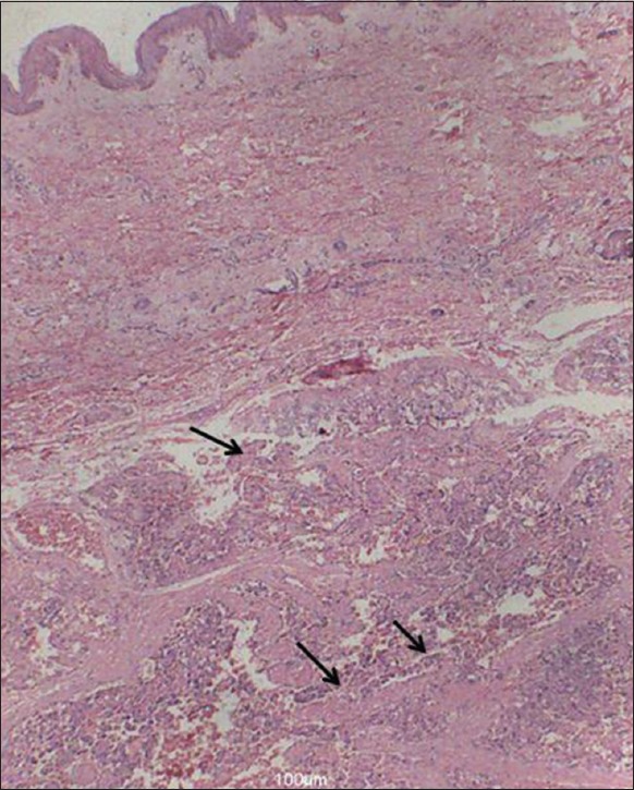
The lesion is well-circumscribed with multiple small papillary structures (arrows) and abutting to the papillae are dilated blood vessels (cavernous hemangioma). Overlying epithelium is also seen (H and E, ×100)

The lesion is well-circumscribed with multiple small papillary structures (arrows) and abutting to the papillae are dilated blood vessels (cavernous hemangioma). Overlying epithelium is also seen (H and E, ×100)