
Fig. 4.
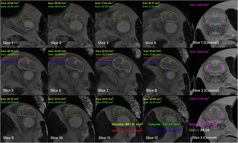
The brain segment result of a chick embryo at 17 days of incubation slice by slice in sagittal and coronal planes. The area of the whole brain (yellow), telencephalon (green), cerebellum (red), brainstem (blue) and LV (pink) were calculated by the ImageJ automatically. Since the slice thickness is 1 mm, the volume is the sum of each slice’s area, which is shown in the bottom of the figure