
Figure 6.
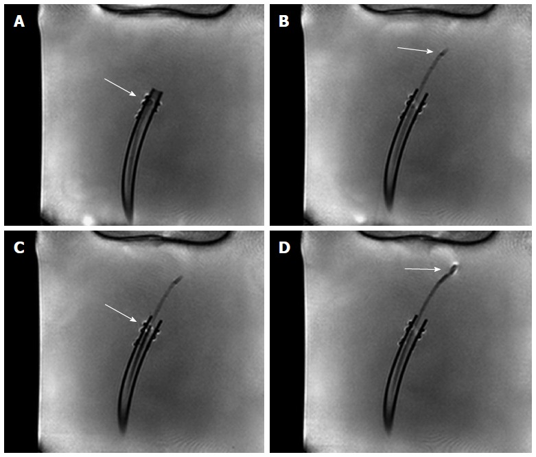
Real-time magnetic resonance imaging image frames showing bioptome and sheath inside saline filled phantom. The empty sheath is pushed forward in a plastic tube (A, B), later the bioptome is guided inside the sheath (C) until the tip is bare in the phantom (D).