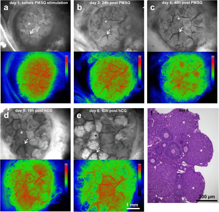
Figure 4. Physiological response of the ovary to hormonal stimulation.
(a–e) Stereomicroscope images and perfusion maps of the mouse ovary undergoing hormonal changes upon injection of pregnant mare’s serum gonadotropin (PMSG; injected at day 2) and human chorionic gonadotropin (hCG; injected 48 h after PMSG, at day 4). Asterix indicates the same ovarian follicle at different time points; arrow denotes the same blood vessel. Calibration bars denote color-coded intensity range (0–255) which corresponds to blood flow map of each time point. (f) Hematoxylin and eosin-stained histological section of the ovary showing corpora lutei formed after ovulation. Asterix denote corpora lutea. a-e were imaged in-vivo in the imaging window.

