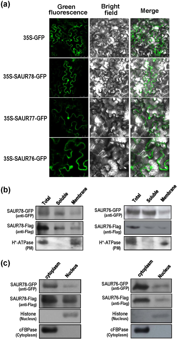
Figure 4. Subcellular localization of SAUR76-78 proteins.
(a) Confocol images of SAUR-GFP proteins transiently expressed in tobacco leaves. (b) Fractionation analysis of SAUR-GFP and SAUR-Flag proteins in transgenic seedlings by Western blot. Presence of SAUR78 (left panel) and SAUR76 (right panel) are shown. H+-ATPase is used as a membrane marker. (c) Subcellular fraction analysis of SAUR-GFP and SAUR-Flag proteins in transgenic seedlings. Presence of SAUR78 (left panel) and SAUR76 (right panel) are shown. Histone H3 and cFBPase are used as nuclear and cytosolic fraction markers, respectively.

