
Fig. 1.
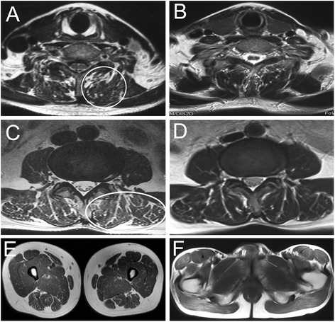
Dynamic changes of muscular MRI. Cervical MRI revealed a few abnormal signals infiltrating into cervical paravertebral muscles in T2 weighted image (a, circle), while the abnormal signals almost disappeared after riboflavin treatment (b). Lumbosacral MRI showed a lot of abnormal signals diffusing around musculi multifidus, musculi longissimus, and iliopsoas in T2 weighted images (c, ellipse), while the abnormal signals almost disappeared after riboflavin treatment (d). Lower limb MRI showed normal pattern of musculi biceps femoris, musculi quadriceps femoris (e), and gluteus maximus (f)